June 2009 Vol. 236 No. 6
Features
Digital Pipeline Radiography Speeds Detection Of Corrosion And Other Anomalies
Recent developments in solid-state radiographic pipeline inspection equipment, primarily durable, mobile computers and solid-state image capturing devices, are raising the technique’s profile as a useful, capable way to find and assess corrosion.
This article discusses how the integration of computers and solid-state imaging systems have increased the power of radiography in inspecting in-service hydrocarbon pipelines.
Radiographic inspection had its beginning with the accidental discovery of X-rays in 1895 by Professor Roentgen, but it was confined to medical usage until the development of high vacuum tubes, first in 1913 and then in 1922. The modern era started in 1931 when GE developed 1MV X-ray generators. At that time, the American Society of Mechanical Engineers granted approval for the use of radiographic images to determine the quality of welded pressure vessels. This led to the acceptance of employing industrial radiography to non-destructively determine weld quality.
Until the last decades of the 20th century, radiography changed little. Although film quality and sensitivities were improved significantly, images were still captured statically on film. Film processing had evolved to an automated state which provided more consistent quality, but the basic processes were the same.
Beginning in the 1980s, the advent of computers and their adaptation as rugged, durable, field-portable machines, coupled with the development of solid-state image capturing devices, have spawned rapid changes in industrial radiography. Images can now be captured on a variety of solid-state devices that accelerate the acquisition to “real-time”.
Software is now available to directly enhance the digital images, send the images anywhere in the world for review and provide archiving opportunities with all the flexibility and capability available with many different data storage and viewing programs. Furthermore, technological advances have led to development of smaller, lighter, more portable X-ray equipment and even field-portable linear accelerators used to generate extremely short wavelength, highly penetrating radiation capable of testing thicknesses of materials that could not be efficiently inspected until recently. Gamma ray inspection has also become increasingly capable, with many man-made isotopes that are stronger than naturally occurring radioactive materials and offer a wide range of energy levels and half-lives.
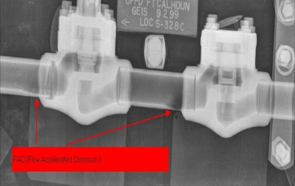
[inline:integritymgmt.JPG=Shown here is the resulting image from the digital radiography of the pipe elbow pictured in the photograph. The x-ray image reveals pitting that would not be as easily detected using other NDT techniques. Photo courtesy of John Cope, Envision Product Design, LLC and Joseph Galbraith, BP America Production.]
Image Capture
X-ray film is a photographic media that consists of a radiation-sensitive emulsion coated on one or both sides of a transparent substrate. Originally, glass plates were used as a substrate, but during WWI were replaced first with cellulose nitrate and later with a cellulose triacetate base. In the 1960s stronger, more stable polyester came into use as the film base. Since the development of radiography in the 19th century, the emulsions have been composed of a form of gelatin and a silver halide, typically silver bromide with a small quantity of iodide. While photographic film is generally only coated with the emulsion on one side of the substrate, typically X-ray film is coated on both sides.
Traditional films are being replaced today by a variety of digital sensors, which, in general, offer a number of advantages, including a wider dynamic range, and a higher sensitivity to radiation which decreases the radiation dose required, allows for shorter exposure times and reduces the safety area. Furthermore, these devices directly create digital images which enable the use of digital image enhancement and on-site interpretation of the results, and finally eliminate the use of chemicals required in conventional film development.
Image Enhancement And Analysis
Image enhancement and processing software is now readily available and provides interpretive tools that are much more powerful and easier to use than have been available when extracting information from radiographic film. Digital image processing provides tools for 1) improving the quality of the image and thereby increasing the probability of detecting the features of interest, and 2) providing tools that greatly assist the analyst in extracting information for the image.
These include contrast improvement and brightness adjustments that enhance the ability to detect very fine variations in grey levels, low-pass and high-pass filters that deal with signal noise, and measurement tools such as histograms and point-processing algorithms to ascertain the grey-level values or density of regions of interest and the dimensional measurements of objects of interest. In the more sophisticated applications, pattern recognition and automated processing can be achieved.
Analyzing radiographic images digitally not only brings the power of commonly available image enhancement software techniques into play, but also provides simplified sharing, storing and retrieving of the images and the information extracted from the evaluation of the images. Specific image formats have been developed in the industry that avoid loss of image quality, since use of the common compressed image formats such as JPG and GIF can result in irreversible alteration of the image data.
Digital Radiography In Pipeline Inspection
Radiography has been used for inspection of weld quality in pipes since sources of radiation strong enough to fully penetrate metal objects became available in the early part of the 20th century. Any anomaly that creates a change in the amount of radiation that passes through the object can potentially be imaged in radiographs. This includes not only slag, porosity, cracks and other weld defects, but also areas of metal wastage caused by corrosion and buildups of scale or transported products.
Industrial radiographic inspection using film is a batch process; relatively small areas are inspected in each exposure. In situations where radiography is employed in pipe mills to assess the quality of production welds, an early version of filmless radiography called fluoroscopy was used.
Later, in the 1980s technology developed in the medical industry that combined image intensification with fluorescent screens and analog cameras was adapted to field inspection of in-service pipelines, thereby, creating a system capable of cost-effectively inspecting long lengths of a pipeline to locate and evaluate areas of localized corrosion. However, due to the cost of these systems and the size, weight and complexities of the components, their use has been restricted.
These early developments have been supplanted by modern systems that incorporate digital sensors with field-durable electronics and computers and provide rapid acquisition, processing and evaluation of images obtained over much longer lengths of pipe than could be cost-effectively inspected using conventional film-based radiography.
These systems can be deployed similar to conventional film by using computed radiography, which offers advantages of shorter exposure times, higher dynamic range, elimination of both film and chemicals, direct creation of digital images, and the opportunity to use lower doses of radiation which translate directly into smaller safety zones and less disruption of other activities in the vicinity of the inspection work.
Other types of sensors can be deployed remotely by pipeline crawlers, creating the benefits of rapid, real-time imaging of long lengths of pipe with the power of digital image acquisition and processing.
Conclusion
Radiography can provide the most cost-effective non-destructive testing (NDT) technique for locating and evaluating anomalies that can adversely affect the integrity of an operating, in-service pipeline. However, the widespread adoption of the tool has been hindered by the restrictions associated with the use of radioactive sources and the requirements for chemically developing the exposed film in a darkroom environment.
That hindrance has been eliminated today by advances in the sources and the image capturing devices that have rendered the technique faster, more powerful and more cost-effective as a field technique. The systems can be remotely deployed in areas difficult or dangerous to access, and can be used to generate information on an operating pipeline without the preparation such as insulation and/or coating removal required when using other NDT techniques such as ultrasonics.
Not only can digital images of hidden pipe features that can impact its ability to safely operate be directly created, processed, viewed and acted on in real-time in the field, but also the image enhancement software available today can help to quickly determine the safe operating pressure of a damaged pipeline.
Acknowledgement
This article is based on a presentation at NACE International Corrosion/2008 in New Orleans, LA.
Authors
Joseph M. Galbraith and George C. Williamson are with BP America Production Company, Houston, TX.
Michael Creech is with Acuren Materials Engineering and Testing, Houston, TX.
Bibliography
Berthel, A, et al., Digital Radiography: Description and User’s Guide, International Symposium on Digital Industrial Radiology and Computed Tomography, June 2007, Lyon, France.
Galbraith, J.M, et al., Real-Time Radioscopic Inspection of Insulated Piping Systems, NACE Paper 95551, Corrosion/95.
Galbraith, J.M., et al., Through-Wall Radiography to Locate and Evaluate Internal Corrosion in Piping, NACE Paper 07172, Corrosion/07.
Metals Handbook, Volume 11 – Nondestructive Inspection and Quality Control, 8th Edition, American Society for Metals, pages 105-156.

Comments